Table of Contents
Previous Case:
Current Case:
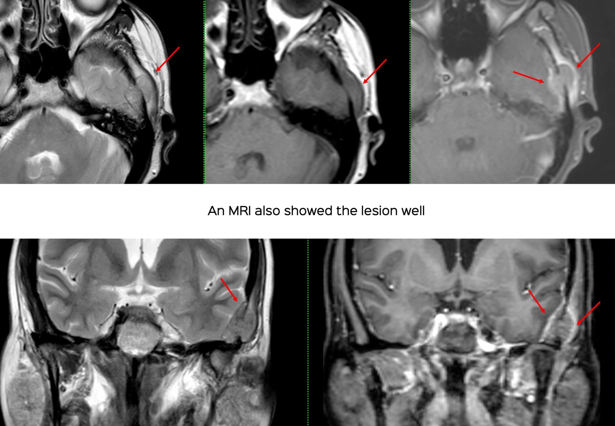
A 59-years old patient diagnosed to have carcinoma rectosigmoid also had a left temporal bone lesion.
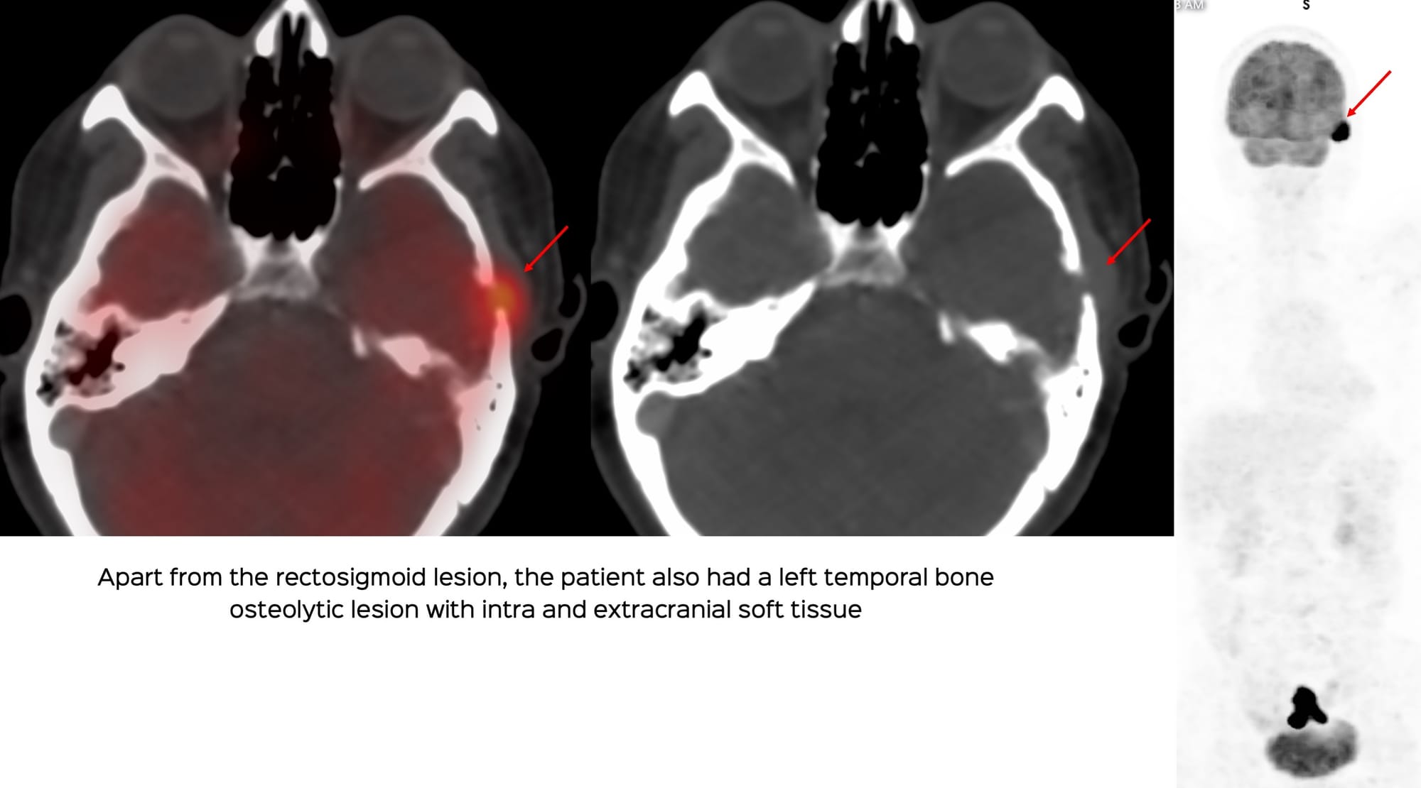
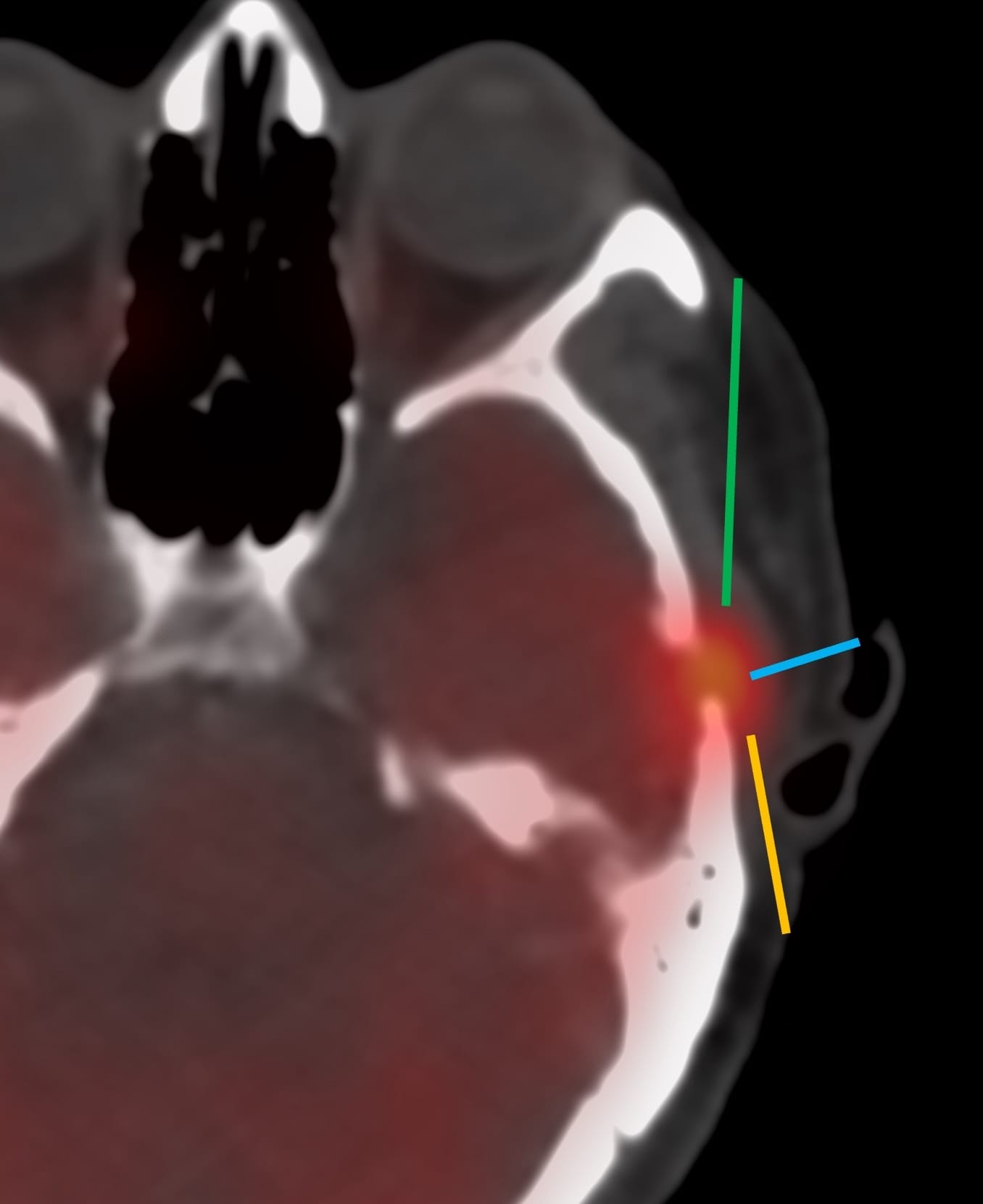
How would you biopsy this lesion?
Please note that the video only plays within this browser page and not outside of this browser page. If the video does not autoplay in your email, then you will need to open this page in your browser, by clicking "View Online", in the 2nd line below the title of the post and next to my name.
Region: Skull, Temporal Bone
Age: 59
Findings: Osteolytic lesion with intra- and extracranial soft tissue
Lesion Biopsied: Left temporal bone lesion
Size of Lesion: 12.4 mm z axis
Gun: 18G BARD, 20 mm throw, but used as 10 mm throw
No of cores: 4 for histopath and aspirate for micro and cytology
Sedation: Yes
Position & Approach: Supine
Time Taken (marker to wash-out): 12 mins
Complication: None
Level of Difficulty: 3/5
Diagnosis: Adenocarcinoma metastasis from carcinoma rectosigmoid
My New Book, including the Kindle Edition
Other Sites and Cases