
Table of Contents
Table of Contents
Table of Contents
 Bhavin Jankharia
Previous Case:
Case 142: Sub 5 mm Lung Nodule Biopsy
Sub 5 mm lung nodules can be biopsied safely by following a few simple principles and steps
Bhavin Jankharia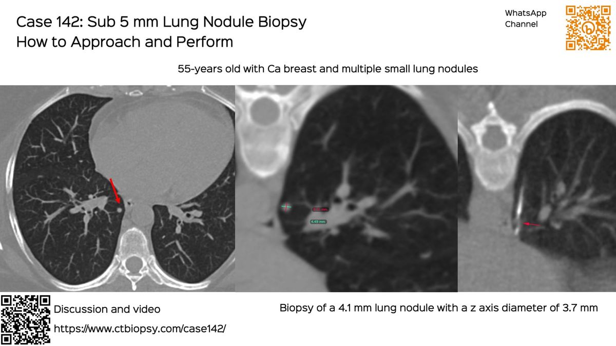
Current Case:
A 71-years old with carcinoma tongue, treated 7 months prior presented with right pleural effusion and small pleural nodules.
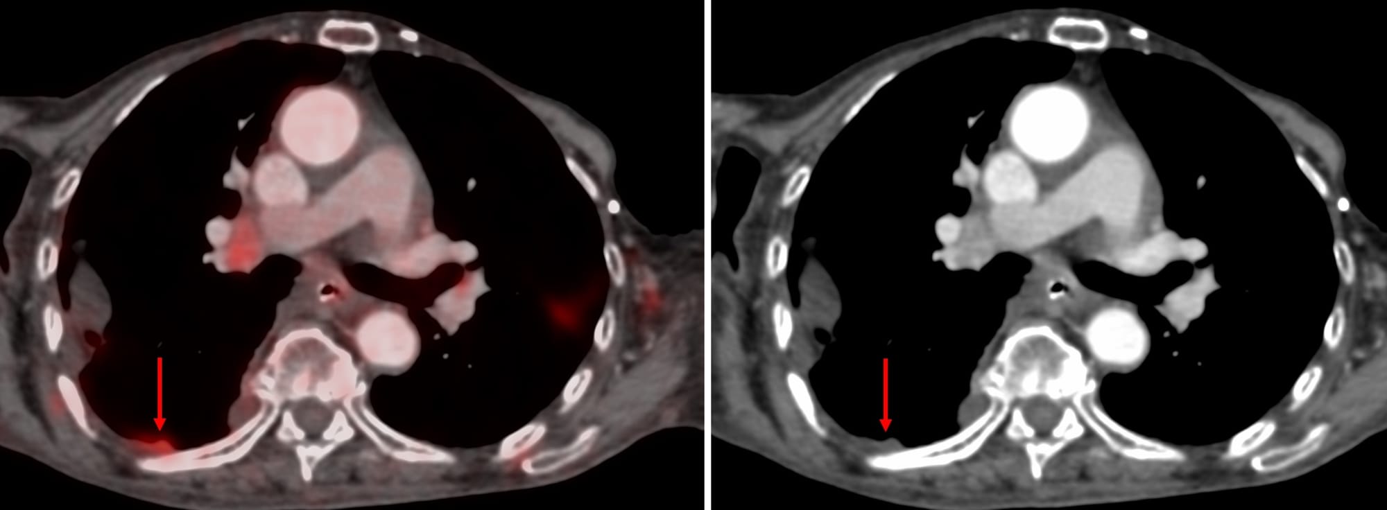
She was referred for a CT guided biopsy. The nodule shown was the most accessible, measuring 2.4 mm in thickness.
There are typically two options - going perpendicular or parallel
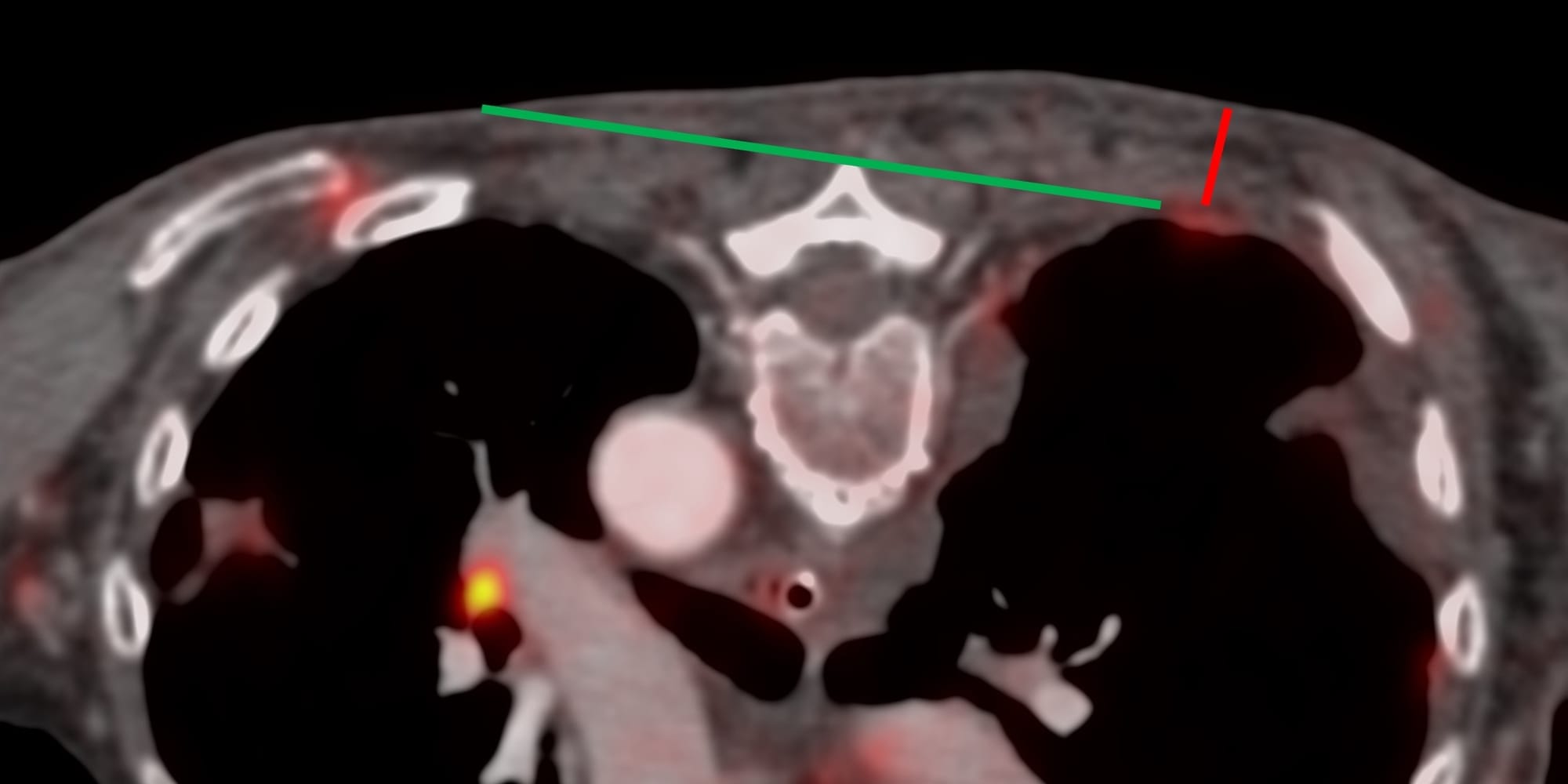
The video discusses the case, the approach to this nodule and basic principles of performing pleural biopsies as far as possible using a parallel approach