Index and Table of Contents
174 Cases with Videos
4 Cases with Images
2 Snippets
5 Lectures
4 Cases of the Day (CODs)

New Onetime Lifetime Subscription

Not all cases need videos. Some can be explained with just images.
Current Case:
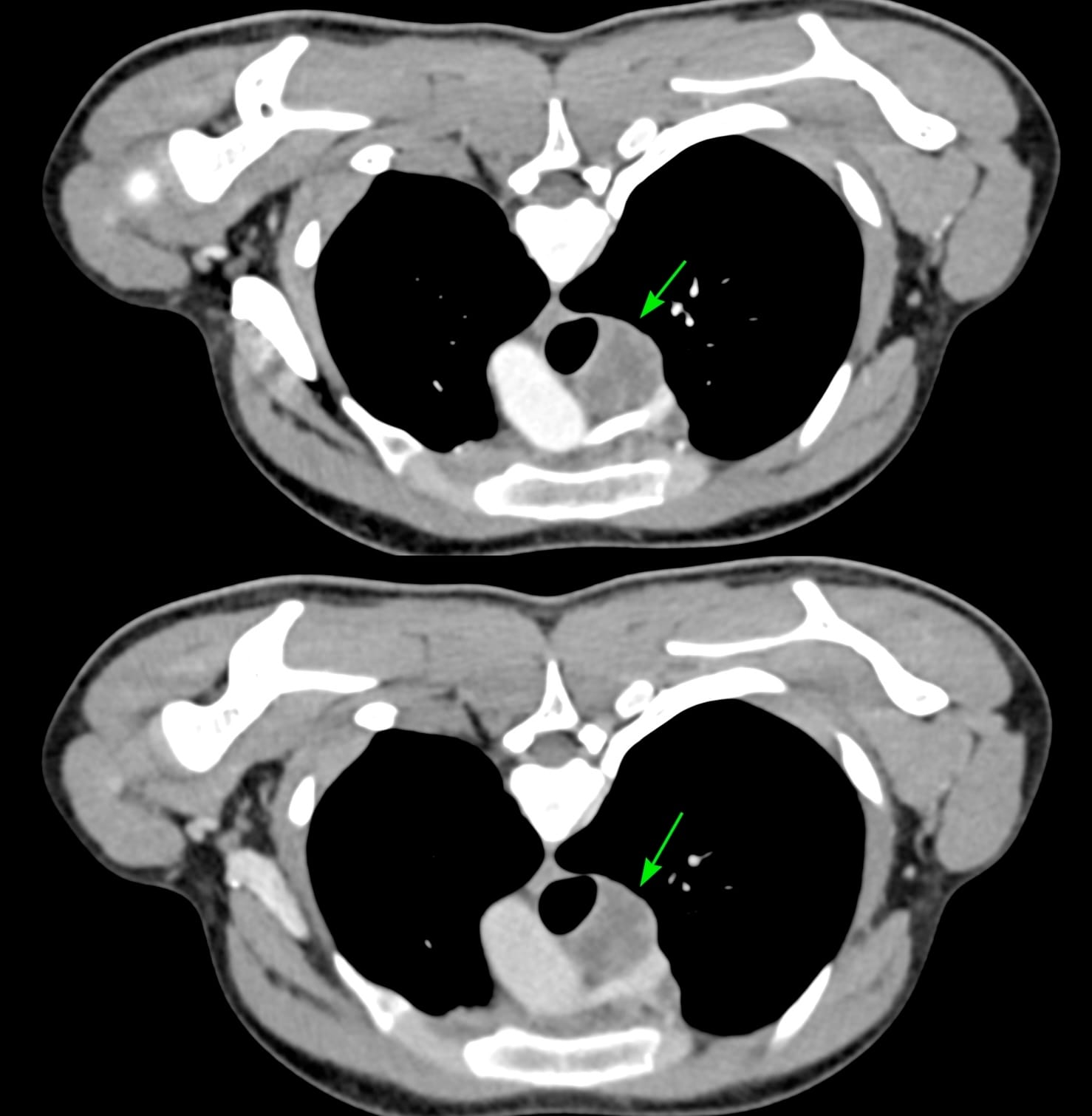
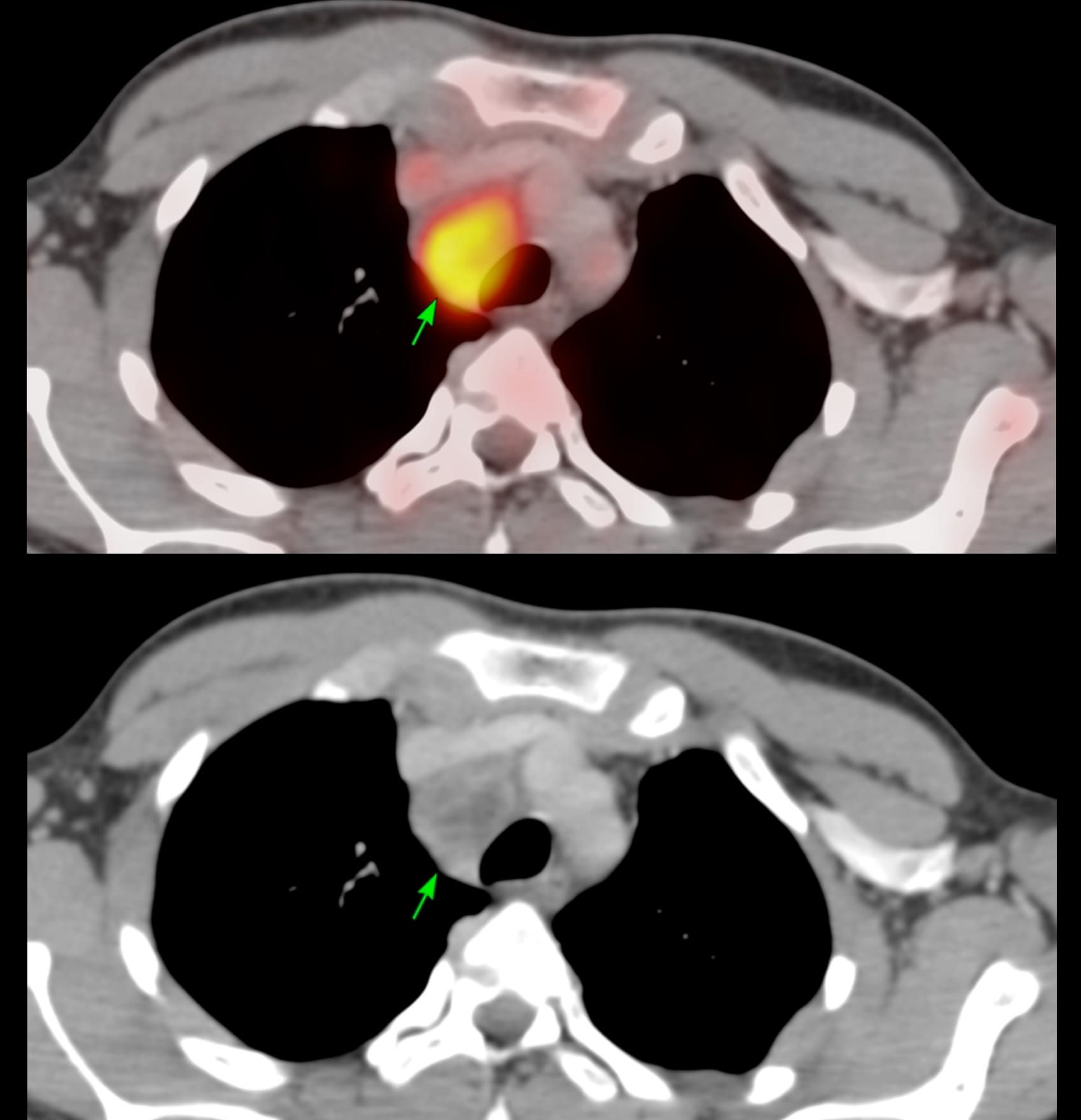
17-years old with fever had a PET/CT that showed a necrotic right paratracheal node - 4R - and was referred for a CT guided biopsy.

The patient was referred for a CT guided biopsy.
Pulmonologists here will jump saying this can be easily done with EBUS, but I have been doing these biopsies under CT guidance since 1995 - over 31 years now, and there is no reason to give them up. EBUS is non-inferior to CT, not better...and it is cheaper, faster, easier and simpler.
Arterial and venous phase studies in the prone position just prior to the biopsy showed the necrotic node well.

While an extrapleural biopsy with hydrodissection is what I usually do, in this case, the lung was extending more medially and a transparenchymal route made more sense.

The next figure shows the procedure. An 18G BARD needle was introduced into the clear space next to the foramen (A) and 10 cc of lignocaine was injected just over the pleura (B). The needle was then passed through a sliver of the lung (C) and then through it into the node (D). 5 cc of pus was aspirated and then intravenous contrast was given before deploying the gun - a hub was used to deploy only 10 mm so as not to overshoot (E). The rapid rollover (F) shows no pneumothorax or hemorrhage. It took 11 minutes.

In the young, it is rare to get a pneumothorax when going through otherwise normal lung and unless you go through a big vessel, hemorrhage is also unlikely. The only thing to take care of is not to overshoot into the mediastinal vessels.
The diagnosis was GeneXpert and histopath positive tuberculosis.
Both extrapleural and transparenchymal routes are part of our armamentarium to access mediastinal lesions and just because EBUS is making inroads does not mean we have to give up doing what was originally the only way to do biopsies, especially since EBUS is not superior to CT guided biopsies - it is just non-inferior and as I said earlier, more expensive and takes much much longer.
Region: Mediastinum
Age: 17
Findings: Necrotic nodes
Lesion Biopsied: Paratracheal node
Size of Lesion: 29.4 mm z axis x 27.7 mm
Gun: 18G BARD, 20 mm throw, long, used 10 mm with hub
No of cores: 3 for histopath and 1 for micro in aspirate for micro
Sedation:
Position & Approach: Prone, transparenchymal
Time Taken (marker to wash-out): 11 mins
Complication: None
Level of Difficulty: 3/5
Diagnosis: Tuberculosis
Similar Biopsy with an Extrapleural Approach

Table of Contents - Other Paratracheal Node Biopsies

Previous Posts:

Other Sites and Cases:
Case of the Day on YouTube

