
Not all cases need videos. Some can be explained with just images.
Current Case:
A 63-years old lady had left ear tinnitus - known to have a cavernous sinus lesion and a carotid space lesion for a few years, indeterminate etiology. A PET showed a lesion in the carotid space encasing the vessels.
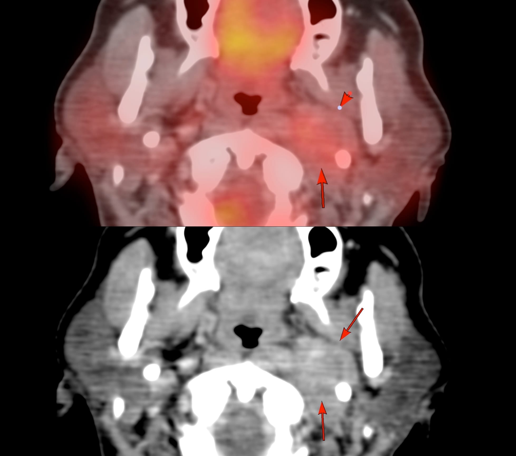
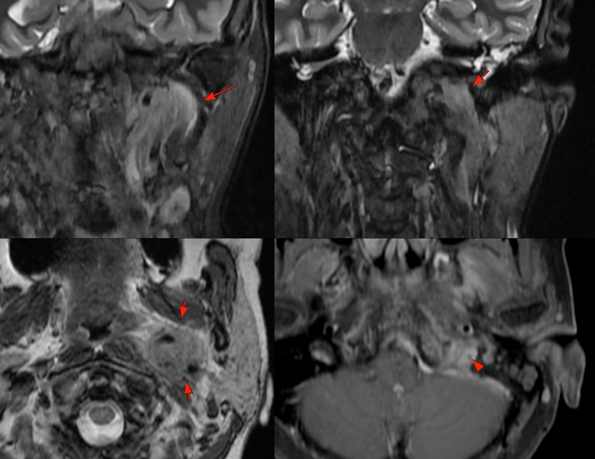
MRI 2 weeks later shows a mass in the carotid space encasing the ICA and extending through the skull base into the dura.
The patient was referred for a CT guided biopsy.
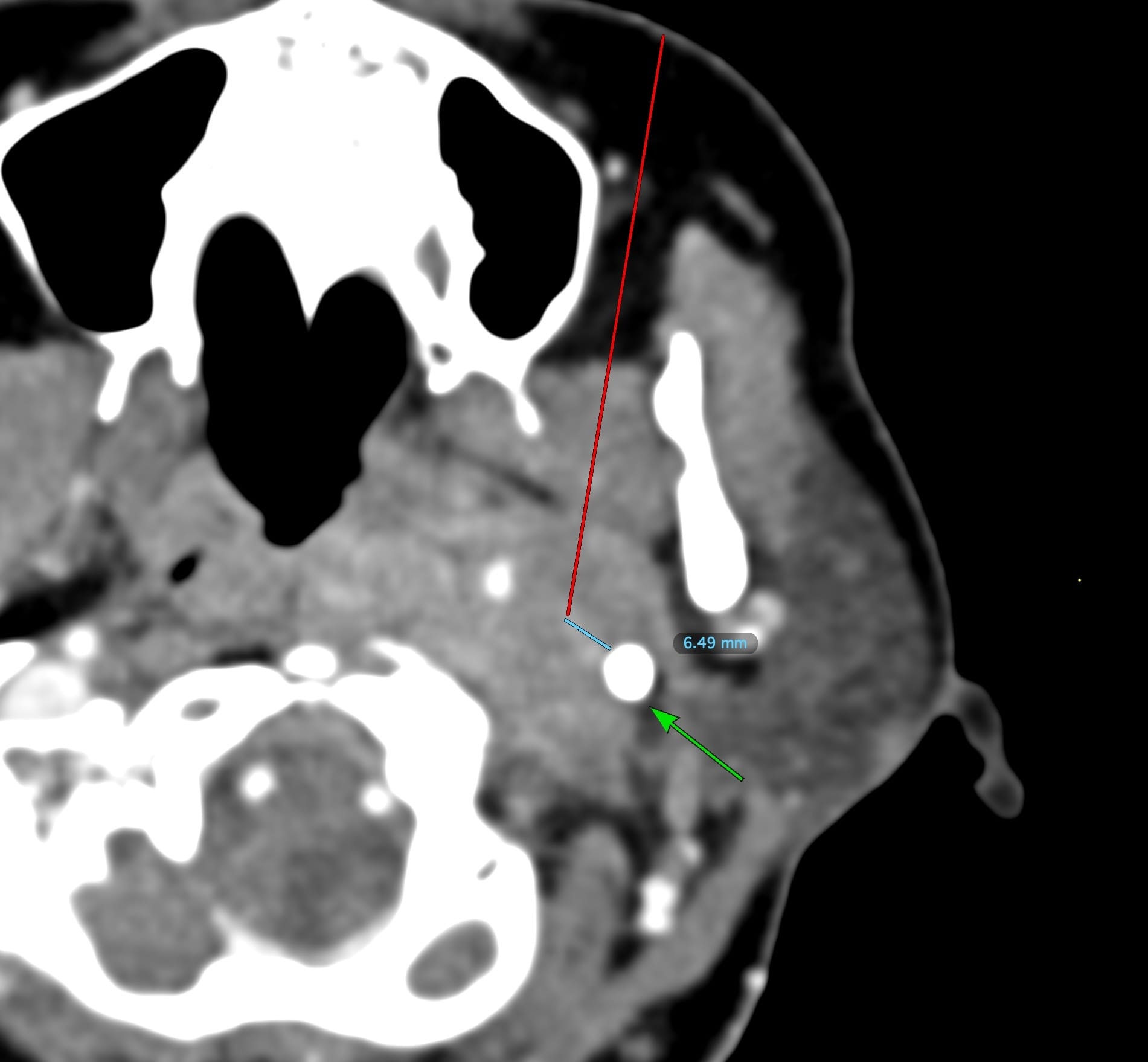
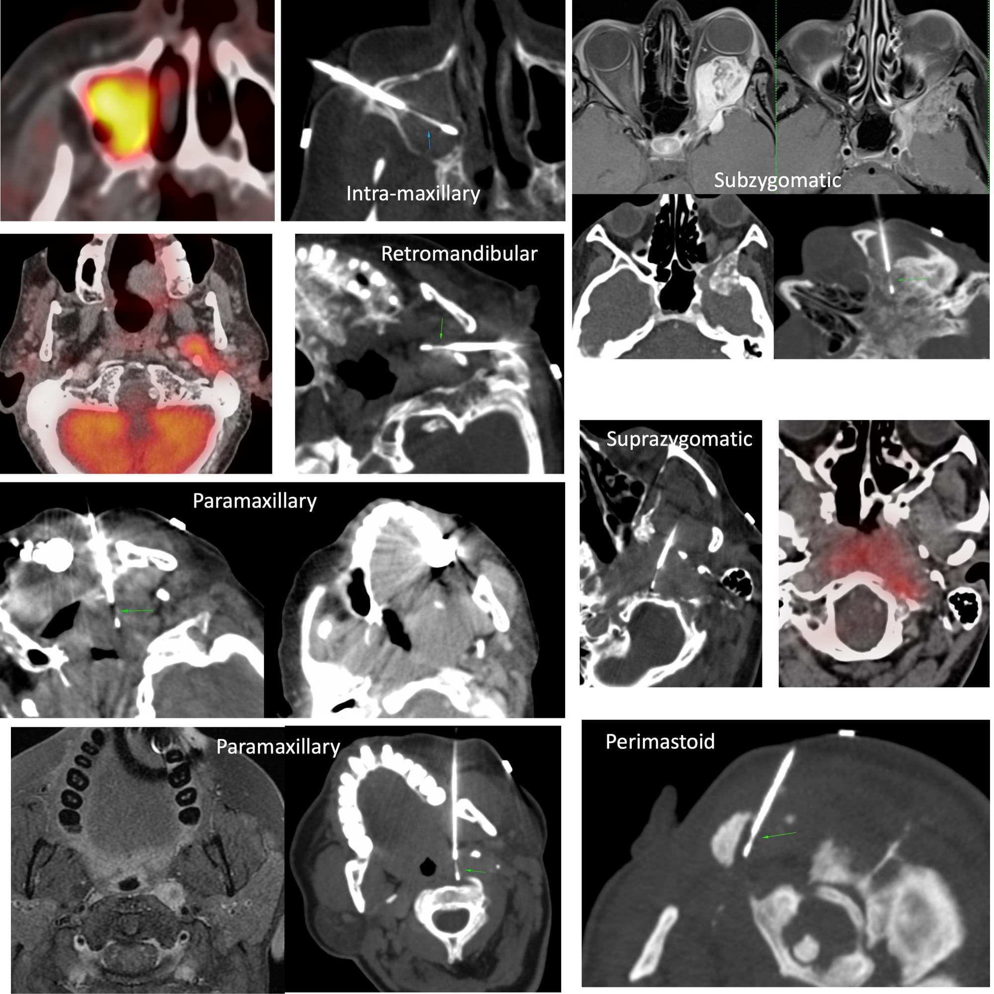
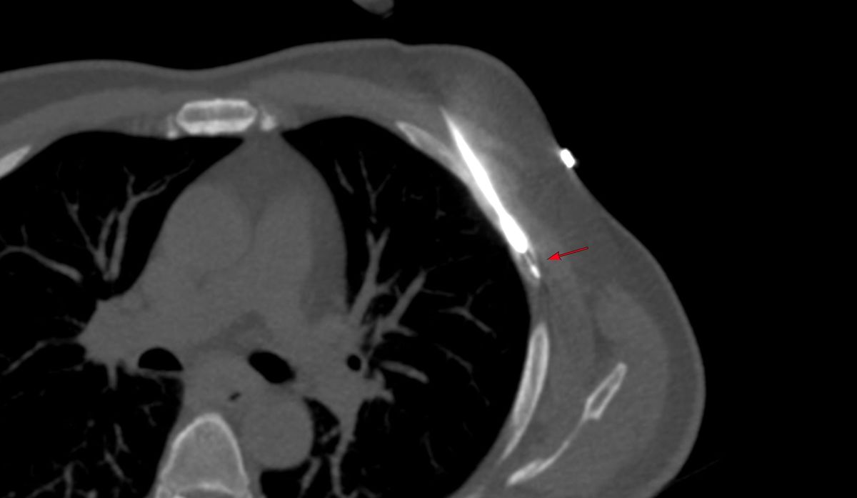
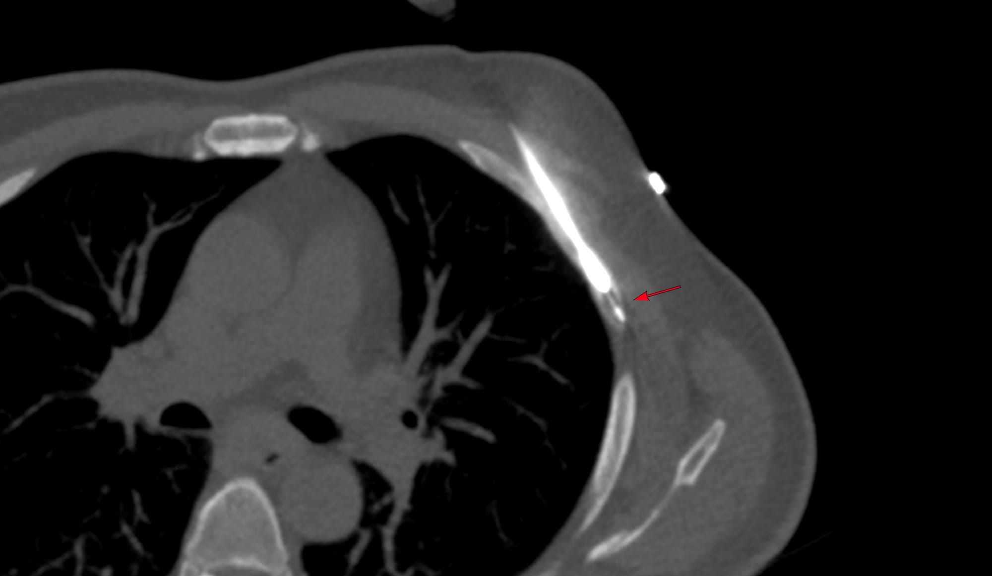
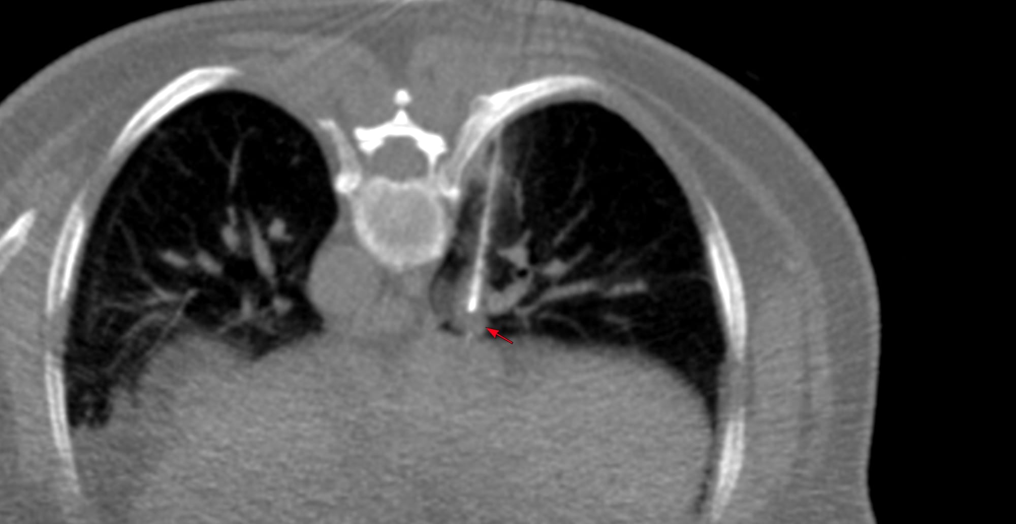
A planning CT angio showed a clear route from the buccal region, past the maxillary sinus, through the pterygoid muscles into the lesion. The idea was to keep the styled process (arrow) as a fixed point marker such that the needle would not be more than 6.5 mm lateral to the process, to avoid the carotid artery.
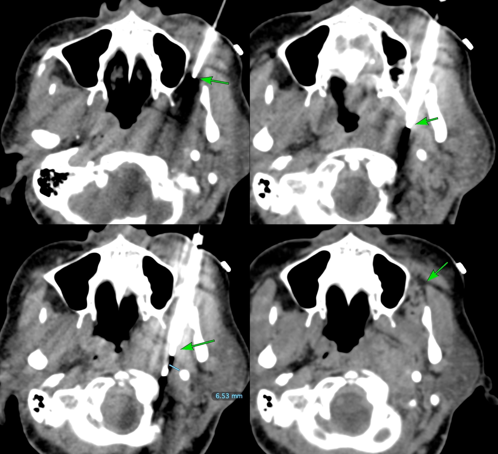
An 18G BARD gun was used to traverse the paramaxillary space and then the pterygoid muscles into the lesion, keeping a safe distance from the carotid, by making sure the needle was under w. A hub was used to convert the 20 mm throw into a 10 mm throw. Three cores were obtained for histology in 12 minutes. There was mild hemorrhage superficially and in the paramaxillary fat, which was not significant.
Region: Head & Neck
Age: 63 years
Findings: Carotid space mass extending up to the skull base
Lesion Biopsied: Carotid space mass
Size of Lesion: 20.4 mm z axis x 24.7 mm
Gun: 18G BARD, 20 mm throw, long, used 10 mm with hub
No of cores: 3 for histopath
Sedation: No
Position & Approach: Supine, transbuccal, paramaxillary
Time Taken (marker to wash-out): 12 mins
Complication: Mild hemorrhage not significant
**Level of Difficulty:**4/5
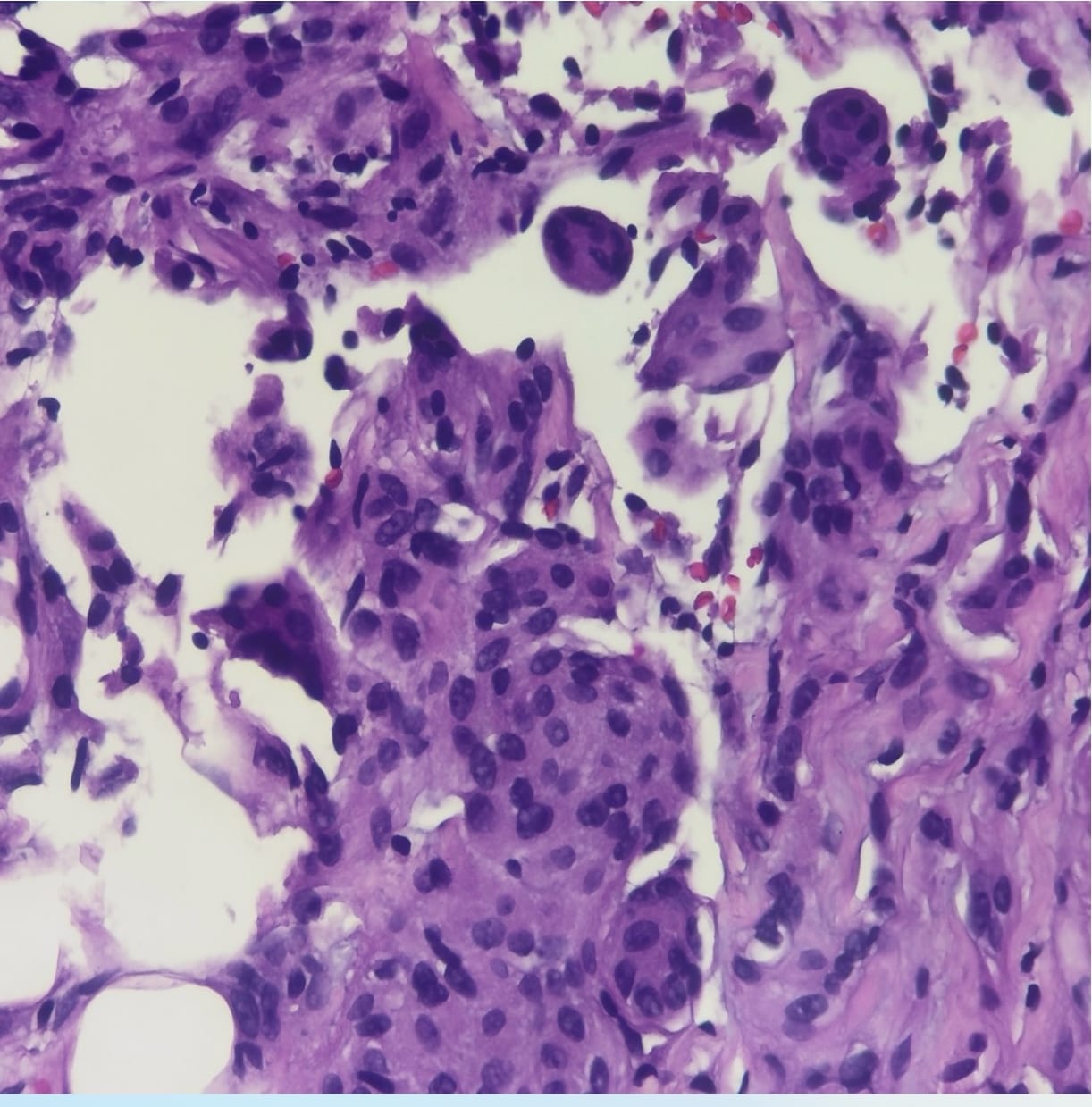
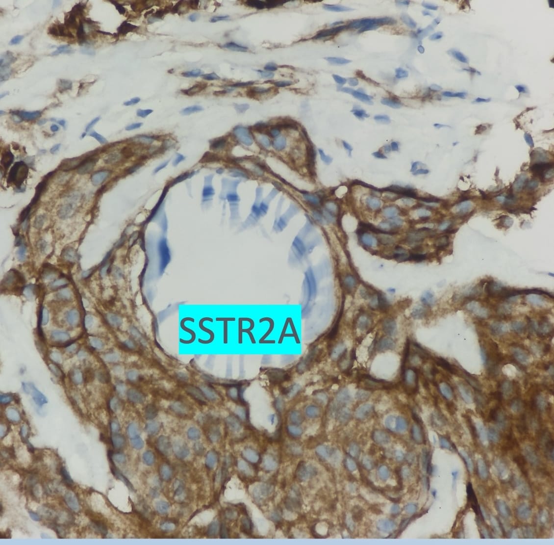
Diagnosis: Meningioma
The final diagnosis was meningioma.
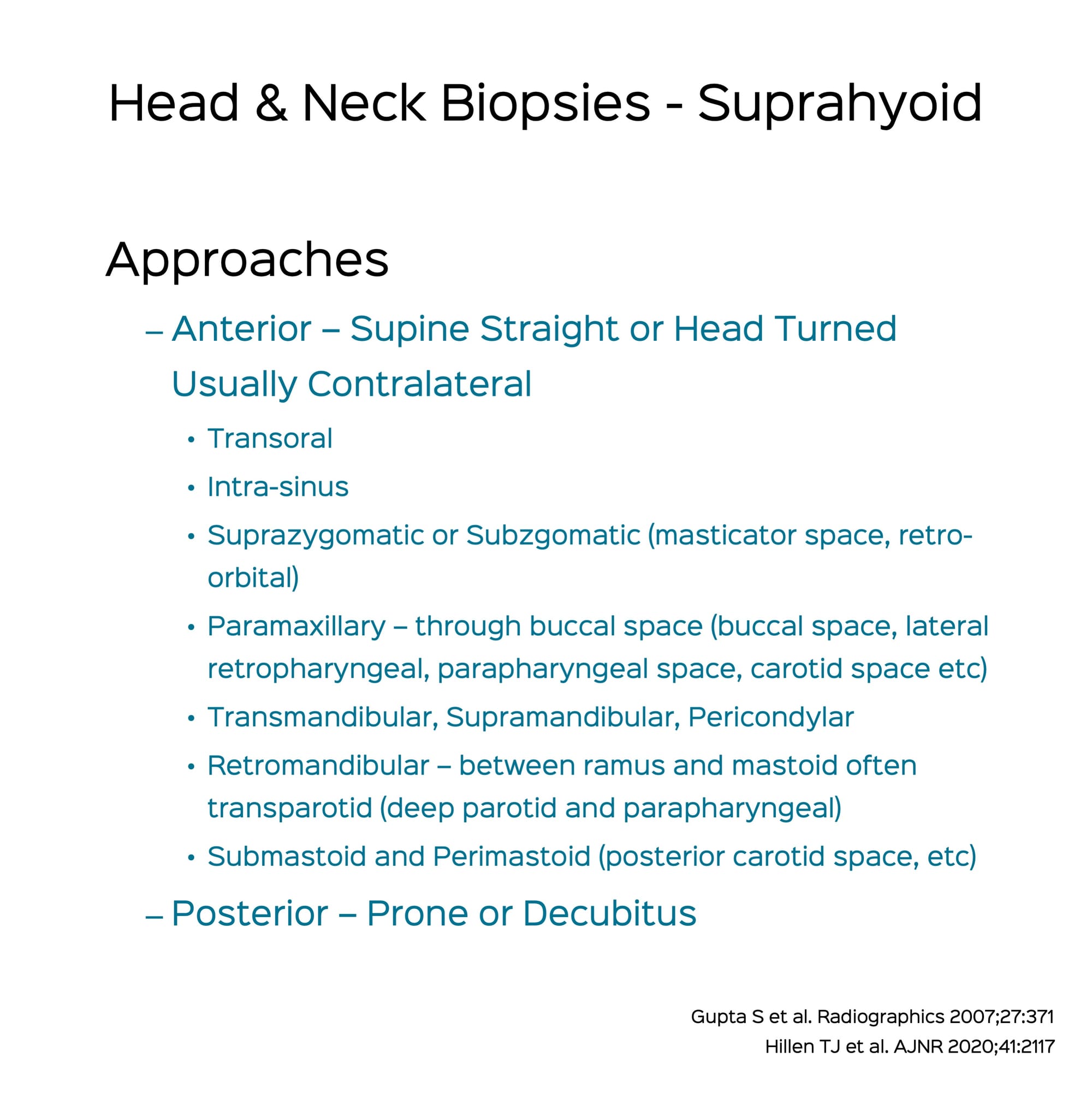
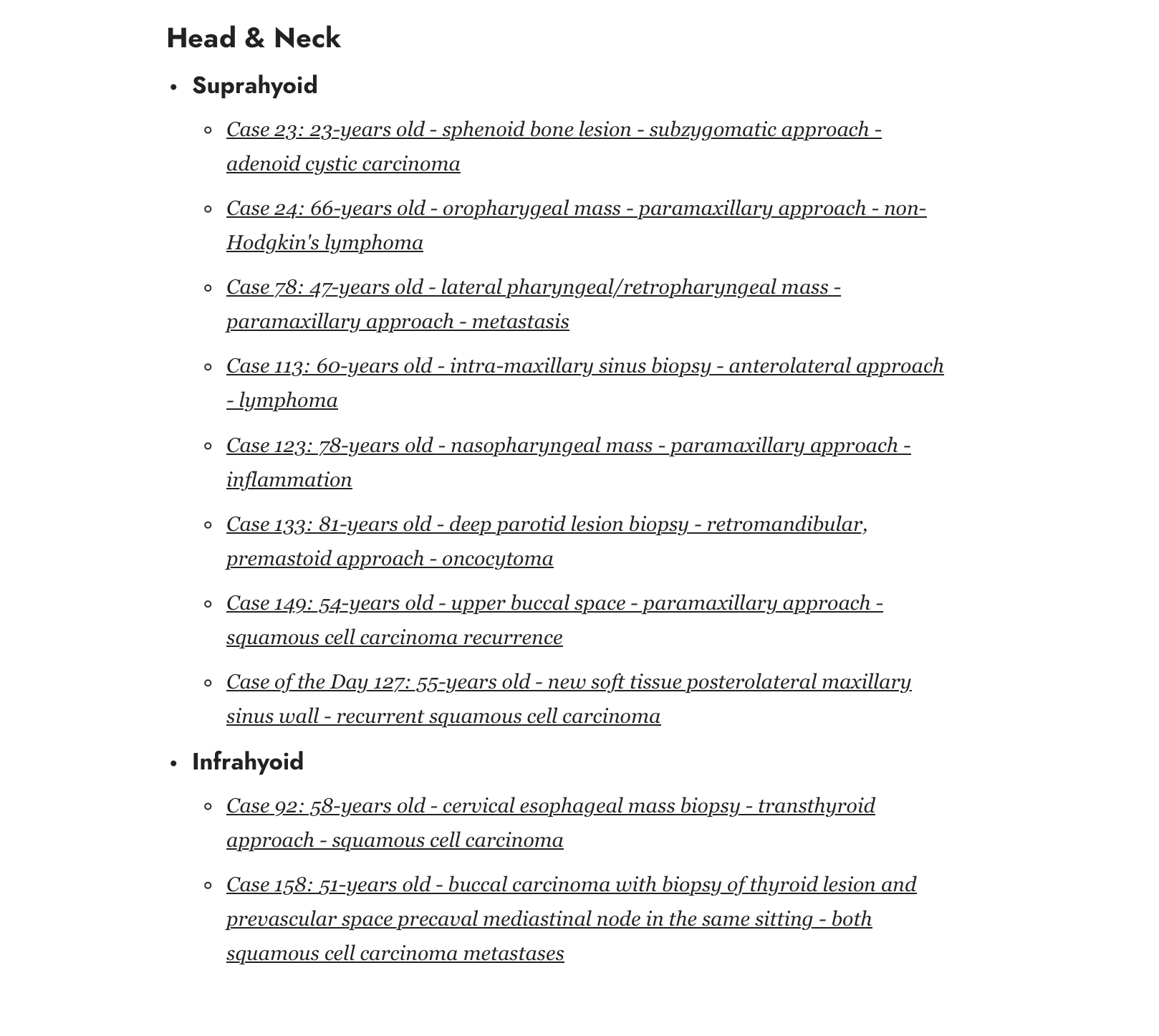
Different CT Guided Biopsy Approaches in the Suprahyoid Neck
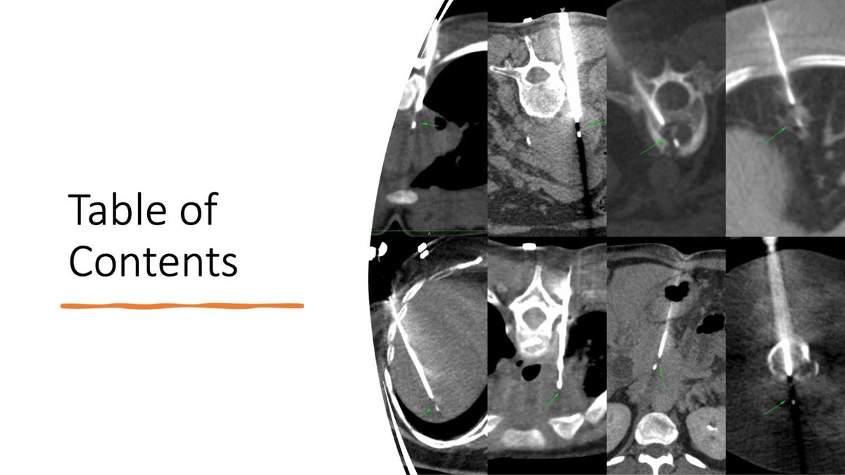
Index and Table of Contents
174 Cases with Videos
7 Cases with Images
2 Snippets
5 Lectures
4 Cases of the Day (CODs)
New Onetime Lifetime Subscription
 Bhavin Jankharia
Table of Contents
Previous Posts:
Other Sites and Cases:
Case of the Day on YouTube