Current Case:
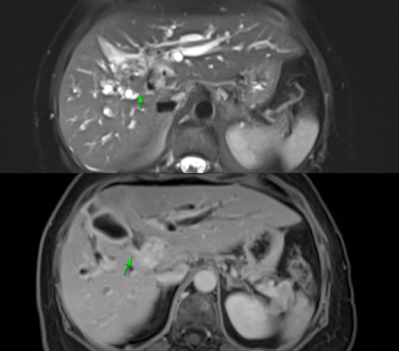
72-years old lady presented with jaundice. MRI (they didn't do an MRCP - cannot you believe it) showed a hilar lesion, likely a cholangiocarcinoma (arrows in Fig. 1).

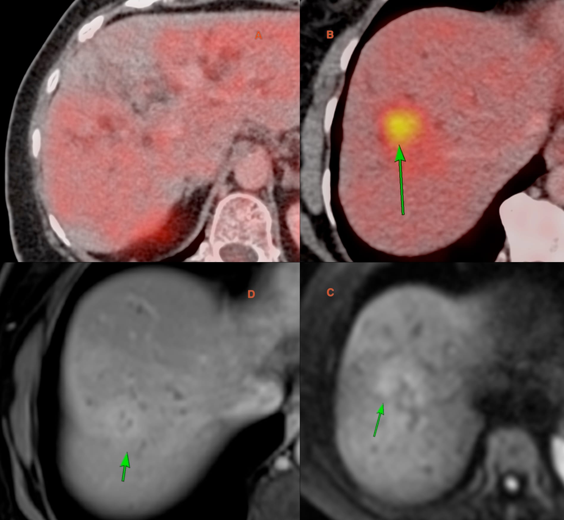
PET-CT (Fig. 2) showed no uptake in the hilar lesion (A), but one lesion in segment 8 (arrow in B) showed uptake. This was retrospectively seen on the MRI as a focal area of restricted diffusion (arrow in C) and enhancement (arrow in D).

She was stented. Endoscopic brush biopsy was negative.
The patient was sent for an image guided biopsy. USG did not show the segment 8 lesion, so she was sent for a CT guided biopsy.
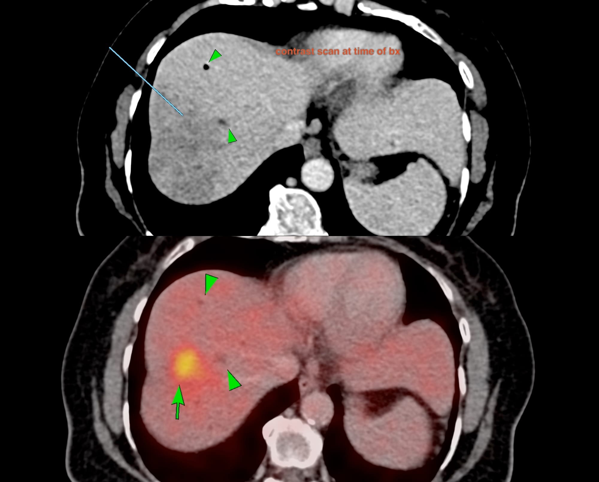
The lesion wasn't well seen on the plain scan, but after giving intravenous contrast, the lesion was seen, ill-defined, in segment 8 (Fig. 3), corresponding to the area of uptake on the PET. I decide to use the two biliary radicles (arrowheads on PET) as my markers (arrowheads on contrast CT). However, the lung was in the way.

Gantry angulation can help. At 10 and 15 deg caudocranial, the lung was just about cleared so finally I settled on 20 deg angulation. As seen, the two biliary radicles were my fixed point markers and there was a clear path to the lesion (Fig. 4).

Fig. 5 and the video show how the biopsy was done. Since the lesion wasn't really seen well and it made no sense to keep giving intravenous contrast, using the biliary radicles as fixed points, the needle was placed lateral to the inferior radicle and then the gun was deployed. The area was gritty, so that was an additional suggestion that the needle was likely in the lesion.

Video
There was no bleeding or complication and the patient left after 90 minutes of observation.
The diagnosis was adenocarcinoma of pancreatibiliary phenotype consistent with an intrahepatic cholangiocarcinoma. Coupled with the imaging, this confirms the extent of the hilar cholangiocarcinoma and the diagnosis.
Region: Liver
Age: 72 years
Findings: Hilar cholangiocarcinoma like lesion with spread to segment 8
Lesion Biopsied: Segment 8 lesion
Size of Lesion: 23.5 mm z axis x 25 mm
Gun: 18G BARD, long, 20 mm throw
No of cores: 3 for histopath
Sedation: No
Position & Approach: Supine, 20 deg caudocranial angulation
Time Taken (marker to wash-out): 13 mins
Complication: None
Level of Difficulty: 4/5
Diagnosis: Intrahepatic cholangicarcinoma
Gantry angulation helps clear vessels, ribs in the chest


and in the liver, especially for segment 8 and 7 lesions with an anterior approach to avoid the lungs. Another trick that helps is after the gantry angulation clears the lungs, to inject 10-20 cc of a saline lignocaine mix at the site of entry to further push the pleura and lung away.
Different CT Guided Liver Lesion Biopsies


Index and Table of Contents
174 Cases with Videos
9 Cases with Images (With or Without Short Videos)
2 Snippets
5 Lectures
4 Cases of the Day (CODs)

New Onetime Lifetime Subscription

Table of Contents

Previous Posts:

Other Sites and Cases:
Case of the Day on YouTube


