New Onetime Lifetime Subscription
Payment
To make the site more accessible, we have decided to remove the yearly subscription and keep only a one-time, lifetime payment to get access to all content at www.ctchestreview.com and www.ctbiopsy.com. These sites are linked and hence one payment gives access to both sites, but the
 Bhavin Jankharia
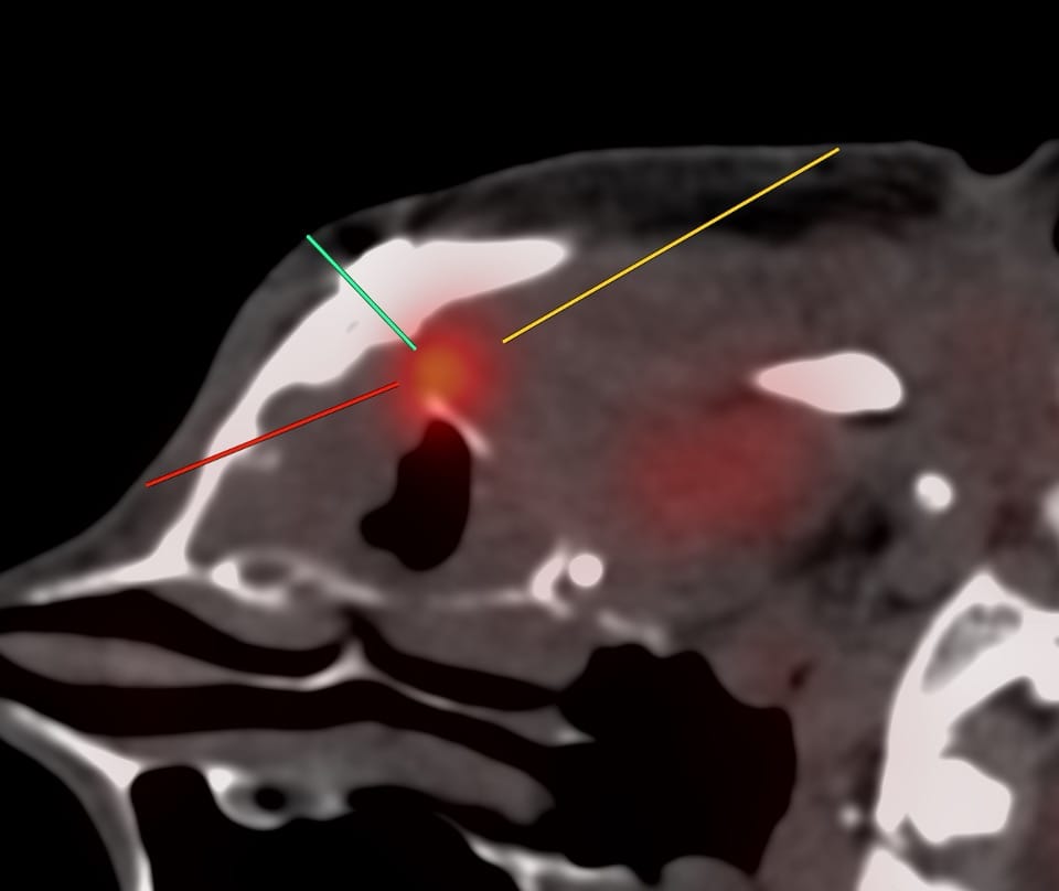
Case:
55-yrs old treated buccal ca with focal soft tissue and uptake on a follow-up PET in the left posterolateral maxillary sinus wall was referred for a CT guided biopsy.
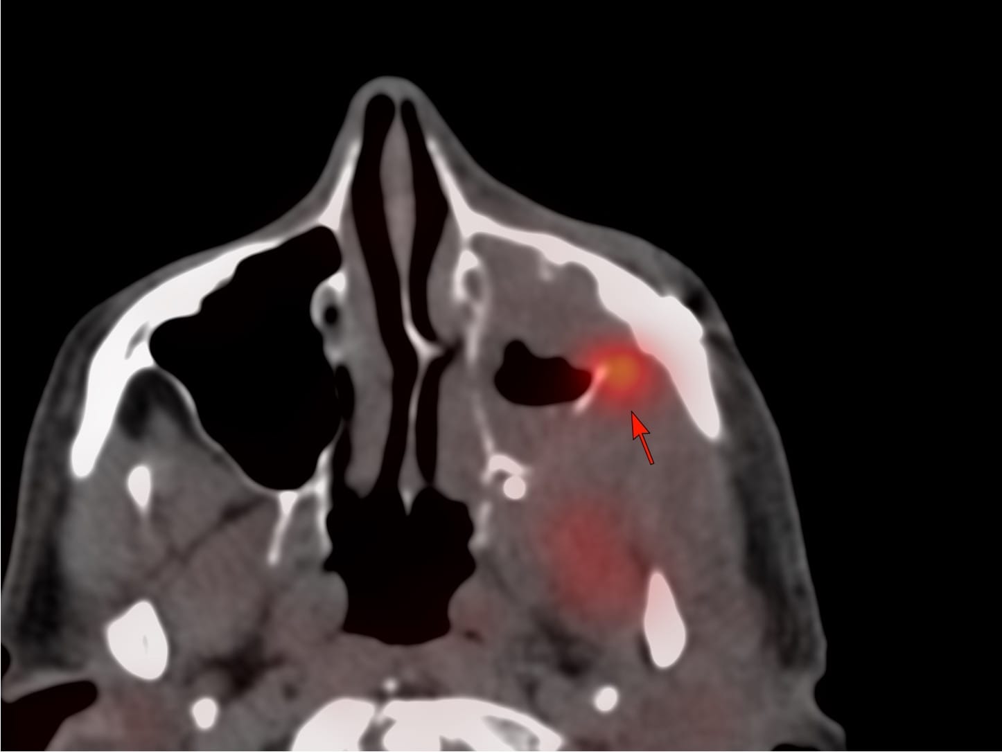
What would be your approaches?
The video discusses the case and describes the procedure and goes through suprahyoid CT guided biopsies in general.