
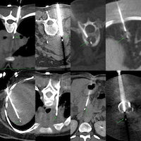
Table of Contents
Table of Contents
Table of Contents
 Bhavin Jankharia
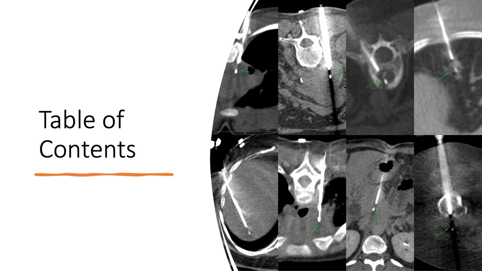
Previous Case:
Case 143: Long Parallel Approach to Small Pleural Lesions
Pleural lesions can be easily biopsied using an approach parallel to the long axis of the lesion
Bhavin Jankharia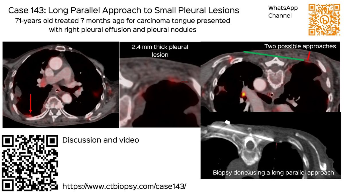
Current Case:
A 77-years old with carcinoma prostate treated 7 years ago, came with a PSMA PET/CT showing a focal sclerotic lesion in the left ischiopubic ramus that had mildly increased in size over 7 years and had an HU of 750.
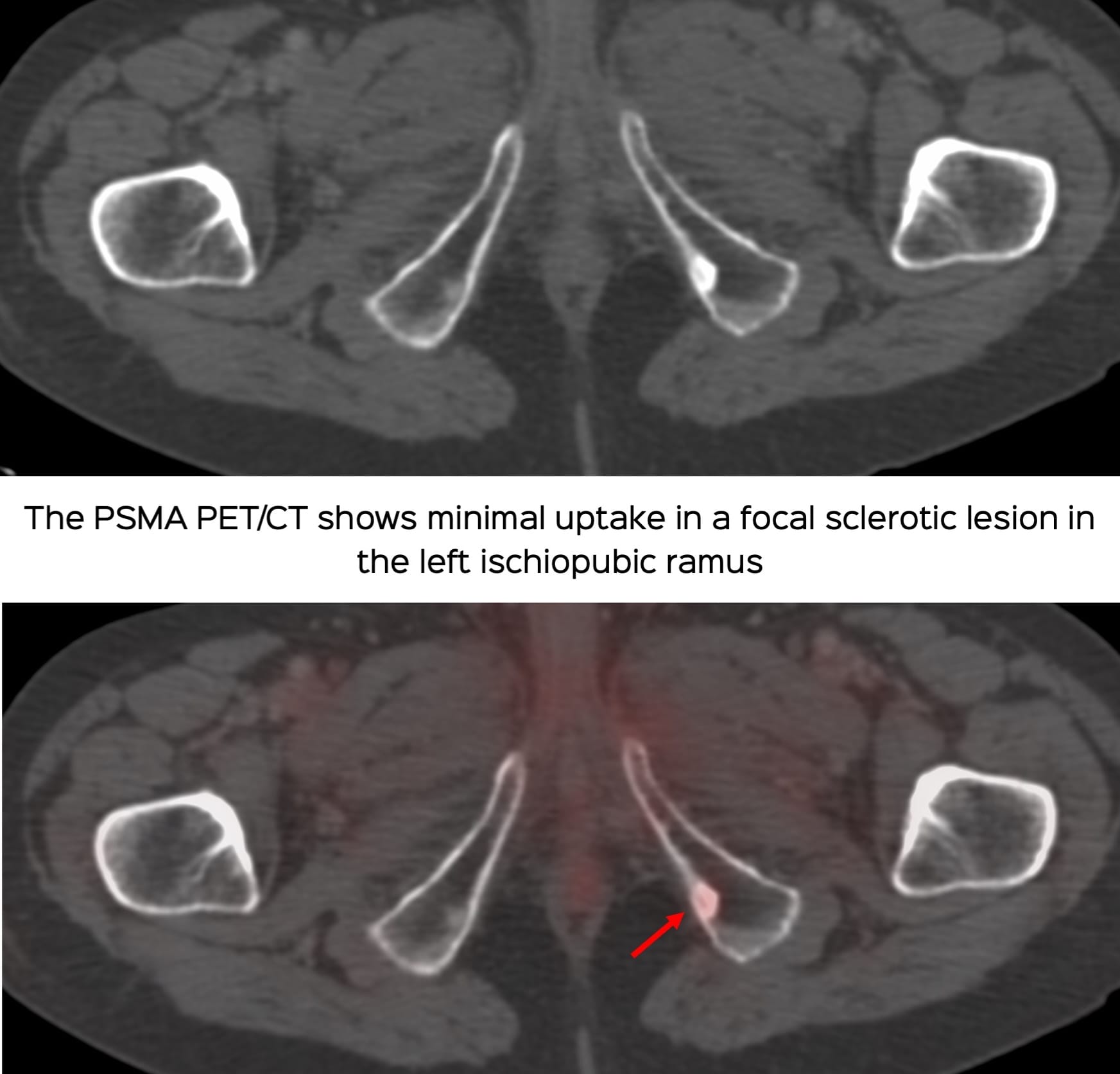
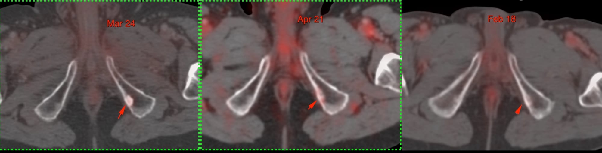
This was the only lesion and hence he was referred for a CT guided biopsy.
The video discusses the case, the approach to this sclerotic lesion and some basic issues with a biopsy of sclerotic bone lesions