New Onetime Lifetime Subscription
Payment
To make the site more accessible, we have decided to remove the yearly subscription and keep only a one-time, lifetime payment to get access to all content at www.ctchestreview.com and www.ctbiopsy.com. These sites are linked and hence one payment gives access to both sites, but the
Current Case:
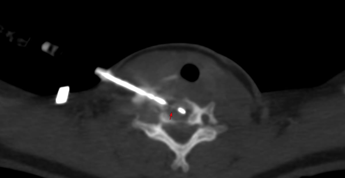
37-years old with radiating upper dorsal pain.
MRI showed a T2 dark lesion involving the D1 vertebral body and pedicle with crenated margins, without trabeculae.
The video discusses the case, the differentials, the final diagnosis with histopath images and one more similar case involving S1.
Region: Spine
Age: 37
Findings: D1 expansile lesion
Lesion Biopsied: D1 lesion - right pedicle
Size of Lesion: -
Gun: 18G Cook, 10 mm throw, long
No of cores: 5 for HP and aspirate for micro and cytology
Sedation: Yes
Position & Approach: Prone - transpedicular
Time Taken (marker to wash-out): 11 mins
Complication: None
Level of Difficulty: 3/5
Diagnosis: Giant cell tumor of bone
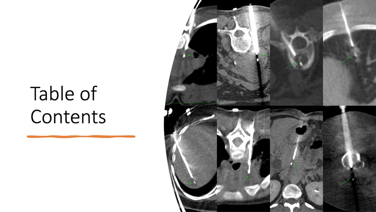
Table of Contents and Other Spine Biopsies:
Table of Contents
Table of Contents
Previous Post:
Case of the Day - 098 - 2025 09 08 - Confirming Tuberculosis of the Lower Cervical Spine
Cervical spine lesions are usually approachable using some route or the other and safe as long as we keep in mind anatomic principles and the positions of the vessels and major nerves.
