Index and Table of Contents
174 Cases with Videos
5 Cases with Images
2 Snippets
5 Lectures
4 Cases of the Day (CODs)

New Onetime Lifetime Subscription

Not all cases need videos. Some can be explained with just images.
Current Case:
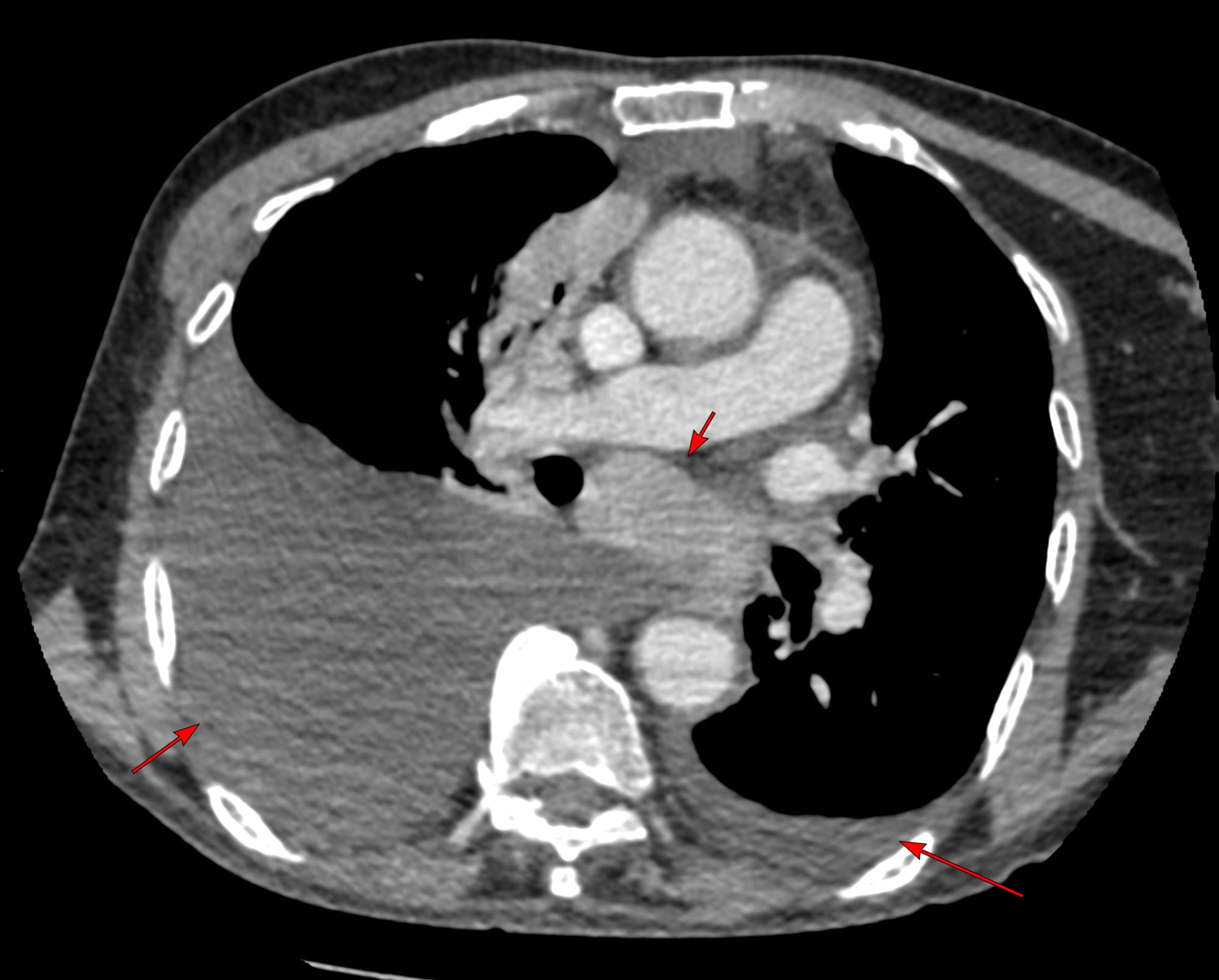
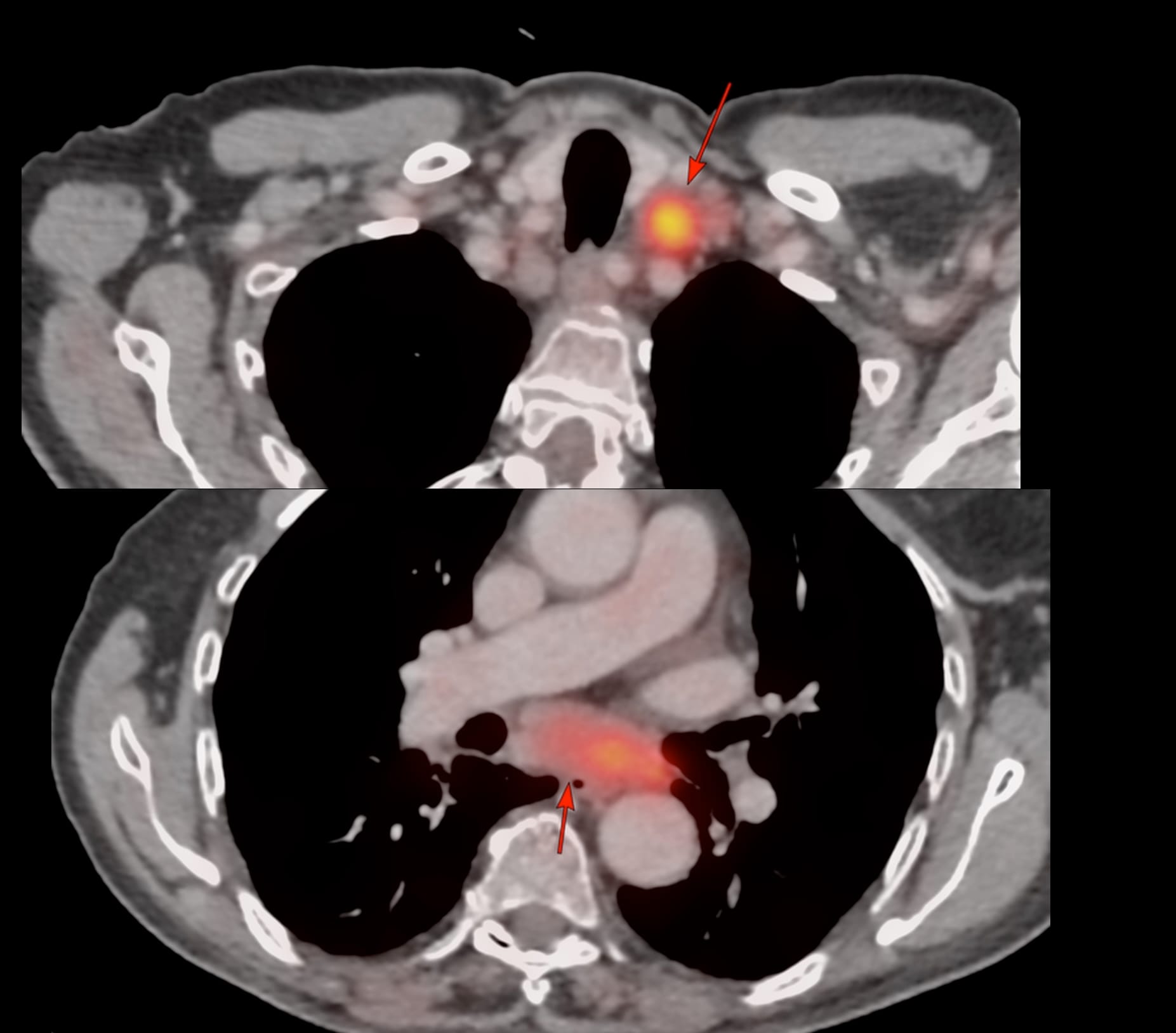
82-years old known to have T-cell non-Hodgkin's lymphoma had a PET in Jan that showed enlarged mediastinal and left supraclavicular nodes.

USG guided left supraclavicular node biopsy was negative for any specific diagnosis.
3 months later, he clinically worsened and CT scan showed the same nodes with mild increase in the subcarinal node size and bilateral effusions and he was referred for a CT guided biopsy of the subcarinal node. The short axis of the node was 25 mm, so biopsy was feasible.

The effusion was a concern, but in the prone position, the fluid moved anteriorly and did not come in the way, though a transeffusion or transpneumothorax biopsy is always a possibility.

The lung was almost at the midline and there was concern that a transparenchymal approach (A) may be needed as in the last case.

Using an 18G Cook needle, 8 cc of lignocaine was injected in the space between the pleura and foramen in the inter-transverse space (B). Once some space was created then it was advanced with 3 injections of 10-15 cc of a combination of lignocaine and saline (C, D), until the needle tip rested along the posterior margin of the node (D) and then it was pushed into the node (E) followed by the biopsy (F). The gun has a 10 mm throw.

6 cores were obtained for histopath and 1 for microbiology and the final diagnosis was relapse of T-cell NHL.
This again takes off my last post on the transparenchymal approach to paratracheal node biopsies. An extrapleural approach with hydrodissection can usually easily be done and while EBUS is commonly used these days, it is not superior to CT guided biopsy, just non-inferior.
Hydrodissection can help push away even lung that extends up to or sometimes across the midline.
This biopsy took 13 minutes from placing the marker to removing the needle and with preparation, counseling, and post-operative stay of 90 minutes, the entire day-care time was 2 1/2 hours and the patient went home normally.
CT guided biopsies are simpler, easier, quicker and much less expensive than EBUS or similar procedures and while fewer and fewer radiologists are learning these skills, they are well worth the effort even today and for the foreseeable future.
Region: Mediastinum
Age: 82
Findings: Enlarged mediastinal nodes
Lesion Biopsied: Subcarinal node
Size of Lesion: 25.1 mm z axis
Gun: 18G Cook, 10 mm throw, long
No of cores: 6 for histopath and 1 for micro
Sedation: No
Position & Approach: Prone, extrapleural
Time Taken (marker to wash-out): 13 mins
Complication: None
Level of Difficulty: 3/5, 4/5
Diagnosis: Relapse of T-cell non-Hodgkin’s lymphoma (NHL)
Similar Biopsies


Table of Contents - Other Subcarinal Node Biopsies

Previous Posts:

Other Sites and Cases:
Case of the Day on YouTube

