Not all cases need videos. Some can be explained with just images.
Current Case:
A 56-year-old man with carcinoma prostate had a PSMA PET (showing uptake in the prostate (A) and in the left 4th rib anteriorly (arrows in coronal MIP and B).

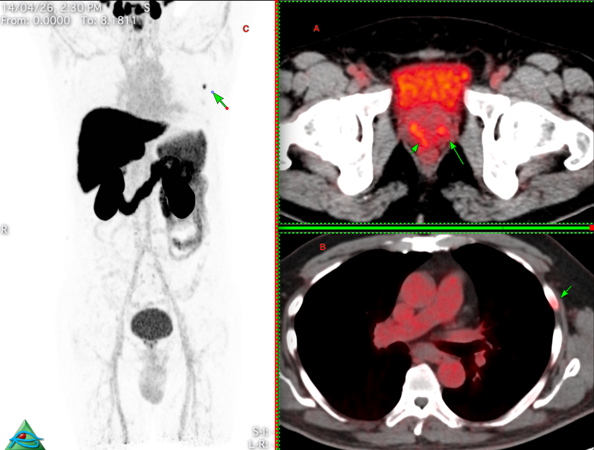
He was sent for an opinion on the rib lesion. In Case 60 at bstneoplasms.com, I discussed a similar case. Most such PSMA-avid rib lesions are benign and incidental — in a recent series by Ou et al. (Skeletal Radiology, 2025), only 5 of 19 biopsied PSMA-avid rib lesions (26%) were metastatic, compared with 82% for spine and 72% for pelvic biopsies.

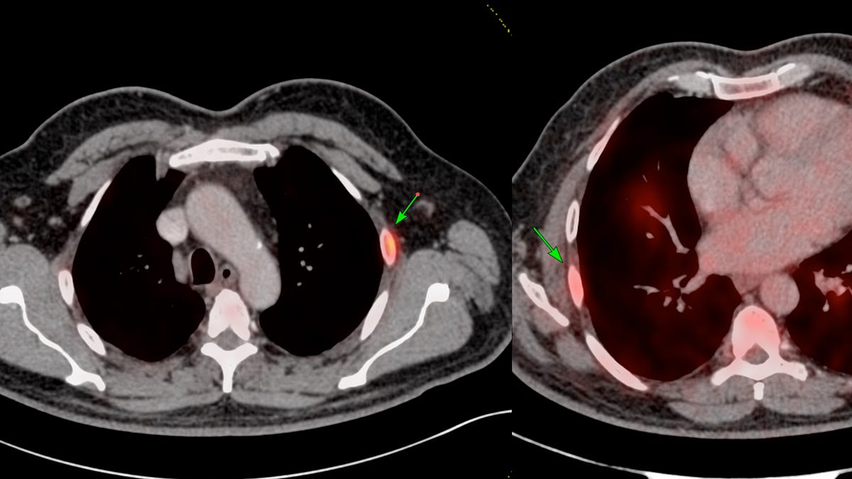
On the bone window and curved MPR images, the lesion had a sclerotic rim without cortical break and no extra-osseous soft tissue — features that favour a benign cyst or fibrous dysplasia. There was no chondroid matrix to suggest enchondroma.
The referring team still wanted tissue.
The transverse route (green line) is tempting — short, straight into the lesion — but the z-axis length is small and the lung is one over-shoot away. The safer choice is to place the needle along the long axis of the rib (blue line). It is technically more demanding, but the throw is controlled and the yield is better.

As the figure below shows, a 13G Cook bone biopsy needle was introduced through the anterior cortex (B) and lightly tapped with a mallet up to the lesion (C). An 18G Cook biopsy gun with a 10 mm throw was passed through it for cores.

Region: Chest Wall
Age: 56 years
Findings: Focal PSMA uptake in a likely benign left 4th rib lesion
Lesion Biopsied: Rib lesion
Size of Lesion: 7.8 mm z axis x 6 mm
Gun: 13G Cook bone biopsy and 18G Cook 10 mm throw, 15 cm long
No of cores: 4 for histopath and aspirate for cytology
Sedation: No
Position & Approach: Supine, along long axis of rib
Time Taken (marker to wash-out): 12 mins
Complication: None
Level of Difficulty: 4/5
Diagnosis: Benign fibrohistiocytic lesion/cyst with hemorrhage
The final diagnosis was a benign fibrohistiocytic lesion/cyst with haemorrhage — as expected.
I covered the full range of rib biopsy approaches in Case 132 at ctbiopsy.com. The two main routes are transverse, which is appropriate for large lesions, large soft tissue components, or when long-axis angulation isn't feasible — and along the long axis, which is the safest and allows multiple cores with minimal pleural or lung injury risk.


Reference:
Ou WC, Jennings JW, Northrup BE, et al. Performance of PSMA-PET/CT as verified by bone biopsy for diagnosing osseous metastases of prostate cancer. Skeletal Radiology. 2025;54(7):1479–1489.
Index and Table of Contents
174 Cases with Videos
6 Cases with Images
2 Snippets
5 Lectures
4 Cases of the Day (CODs)

New Onetime Lifetime Subscription

Table of Contents - Other Subcarinal Node Biopsies

Previous Posts:

Other Sites and Cases:
Case of the Day on YouTube

