
Current Case:
A 62-year-old man with a suspected recurrent lesion around the pterygoid plates on MRI and PET/CT came for a biopsy.
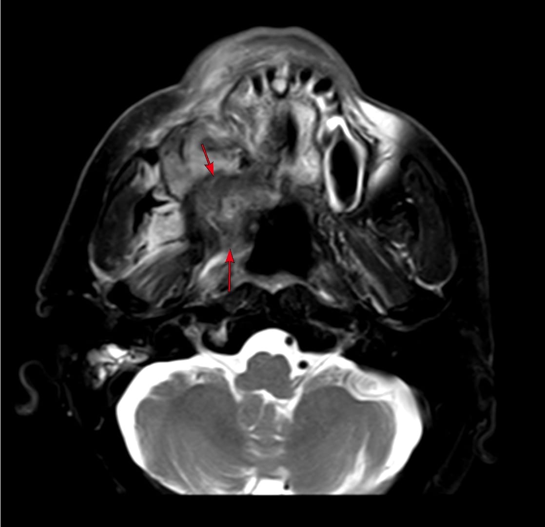
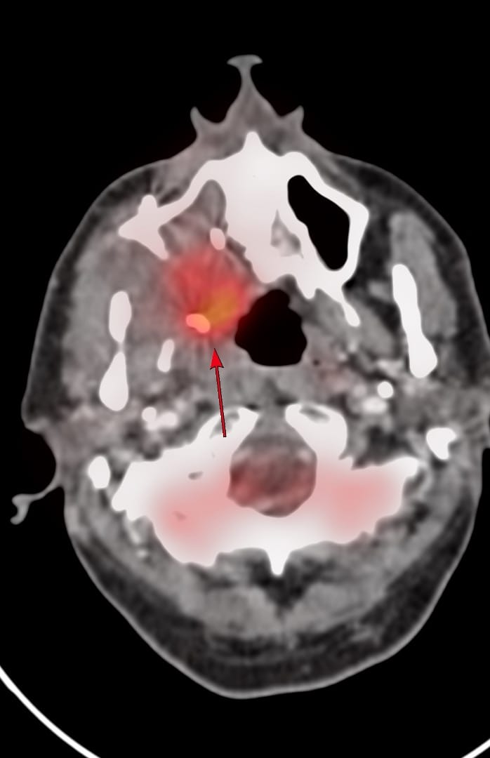
The patient was referred for a CT guided biopsy which was done using a paramaxillary approach.
The video discusses
- The case
- The diagnosis
- The paramaxillary approach referencing a recent AJNR article on this subject.
Video
Region: Head & Neck
Age: 62 years
Findings: Mass along posterior wall of maxillary sinus and peri-pterygoid region
Lesion Biopsied: Same
Size of Lesion: Not relevant
Gun: 18G Cook, 10 mm throw, short
No of cores: 8 for histopath in 4 vials
Sedation: No
Position & Approach: Supine, head mildly turned to left, paramaxillary
Time Taken (marker to wash-out): 8 mins
Complication: None
Level of Difficulty: 3/5
Diagnosis: Squamous cell carcinoma recurrence
Other Cases Using the Paramaxillary Approach
(From superficial to deep)


Index and Table of Contents
175 Cases with Videos
10 Cases with Images (With or Without Short Videos)
2 Snippets
5 Lectures
4 Cases of the Day (CODs)

New Onetime Lifetime Subscription

Previous Posts:

Other Sites and Cases:
Case of the Day on YouTube


